
Not long ago, dental implant planning relied mainly on traditional two-dimensional X-rays. These images were helpful, but they could only show part of the picture. Surgeons had to estimate important details like the width of the bone, the exact location of nerves, and the position of the sinuses.
Today, technology has changed the way implant surgery is planned and performed. Three-dimensional imaging allows oral surgeons to see the jaw from every angle before treatment begins, and guided surgery tools help turn that detailed plan into precise results in the operating room.
For patients, these advances can mean more predictable outcomes, a smoother experience, and greater confidence in their treatment. Here is a closer look at how this technology works and why it matters.
What Is 3D Imaging?
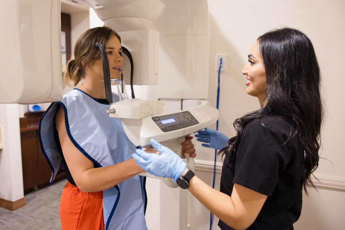
Most 3D dental imaging is done with a cone beam computed tomography scanner, often called a CBCT scan. The scanner rotates around your head and captures a series of images in less than a minute. These images are combined into a detailed three-dimensional model of your teeth, jaw, nerves, and sinuses.
The scan is quick, painless, and non-invasive. You simply sit or stand still while the machine does its work.
What Your Surgeon Can See
A 3D scan gives your oral surgeon information that a traditional X-ray cannot provide. This includes:
- The height, width, and density of the jawbone
- The exact position of nerves and blood vessels
- The location and size of the sinuses
- Any hidden infections, cysts, or abnormalities
- The relationship between the implant site and neighboring teeth
With this information, the surgeon can choose the ideal implant size and position and determine whether a bone graft is needed before surgery ever begins.
Planning the Implant Digitally
Using specialized software, the surgeon can place a virtual implant on the 3D model. This allows them to test different positions and angles and choose the one that offers the best support, function, and appearance.
The plan also considers where the final tooth will sit, so the implant is placed in a position that supports a natural-looking result.
How Guided Surgery Works
Guided surgery helps the surgeon follow the digital plan during the procedure.
In some cases, a custom surgical guide is made from the 3D plan. This guide fits over the teeth or gums and directs the exact position, angle, and depth of the implant.
Other systems use dynamic navigation, which works much like GPS for implant surgery. Cameras and software track the surgical instruments in real time and show the surgeon their position on a screen compared with the digital plan. This allows for precise adjustments throughout the procedure.
Benefits for Patients
Greater precision can make a real difference in the patient experience. Careful planning helps protect nerves and sinuses and supports ideal implant placement.
In many cases, guided techniques allow for smaller incisions, which may help reduce discomfort and swelling after surgery. Knowing exactly what to expect before the procedure can also shorten treatment time and help patients feel more at ease.
3D models can also be used to explain the treatment plan visually, so patients understand what will happen and why.
Is 3D Imaging Safe?
CBCT scans use a focused beam and are designed to capture only the area needed. The radiation dose is generally low compared with a traditional medical CT scan. Your surgeon will only recommend a scan when the information is valuable for your care.
Advanced Care in the Coachella Valley
At Desert Maxillofacial Center, we use 3D imaging and advanced surgical technology to plan dental implant procedures with precision and help our patients feel informed at every step.
We serve patients throughout the Coachella Valley, call (760) 837-1515 to schedule a consultation and learn how modern technology can support your implant treatment.